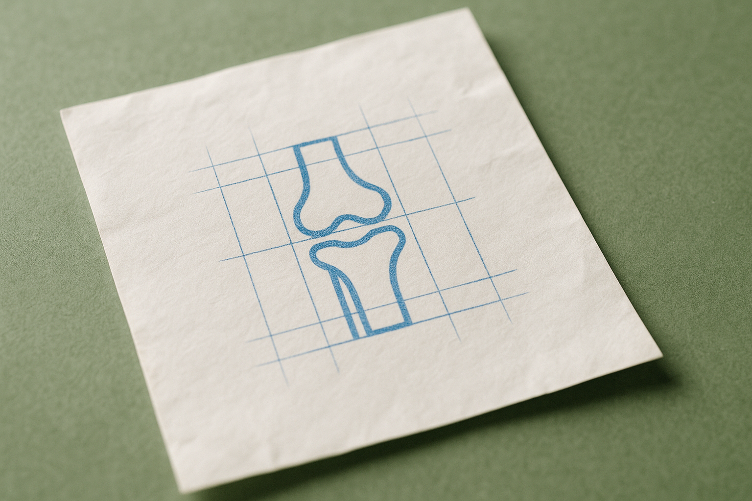
A medical procedure called knee radiofrequency ablation is used to treat long-term knee pain. This treatment involves using a precisely targeted electric current to generate heat, which then interrupts pain signals sent from specific nerves in the knee to the brain.
This publication describes a method for performing this procedure that is tailored to each individual patient. This patient-specific approach uses a detailed, three-dimensional knowledge of the knee's anatomy. Understanding the knee in 3D helps guide the procedure, accounting for the fact that the exact location of nerves can differ between people.
To ensure the treatment is delivered to the correct nerves, a technique called contrast spread analysis is used. Before applying heat, a special dye, known as a contrast agent, is injected. This dye is visible on imaging scans, such as X-rays. By watching how the dye spreads through the tissue, medical professionals can confirm that the instrument is positioned correctly before they begin the nerve ablation.
The information is presented through a series of "teaching images," which are visuals designed to show and explain this specific technique to other healthcare providers.