Chronic lower back pain is sometimes treated with a procedure that uses heat to disable specific nerves that send pain signals from the joints in the spine. This procedure is called lumbar medial branch radiofrequency neurotomy (LMBRFN). However, the bones in the lower back, called lumbar vertebrae, vary in shape from person to person. This anatomical variability can affect the success of the procedure, creating a need for an approach that is customized to each patient.
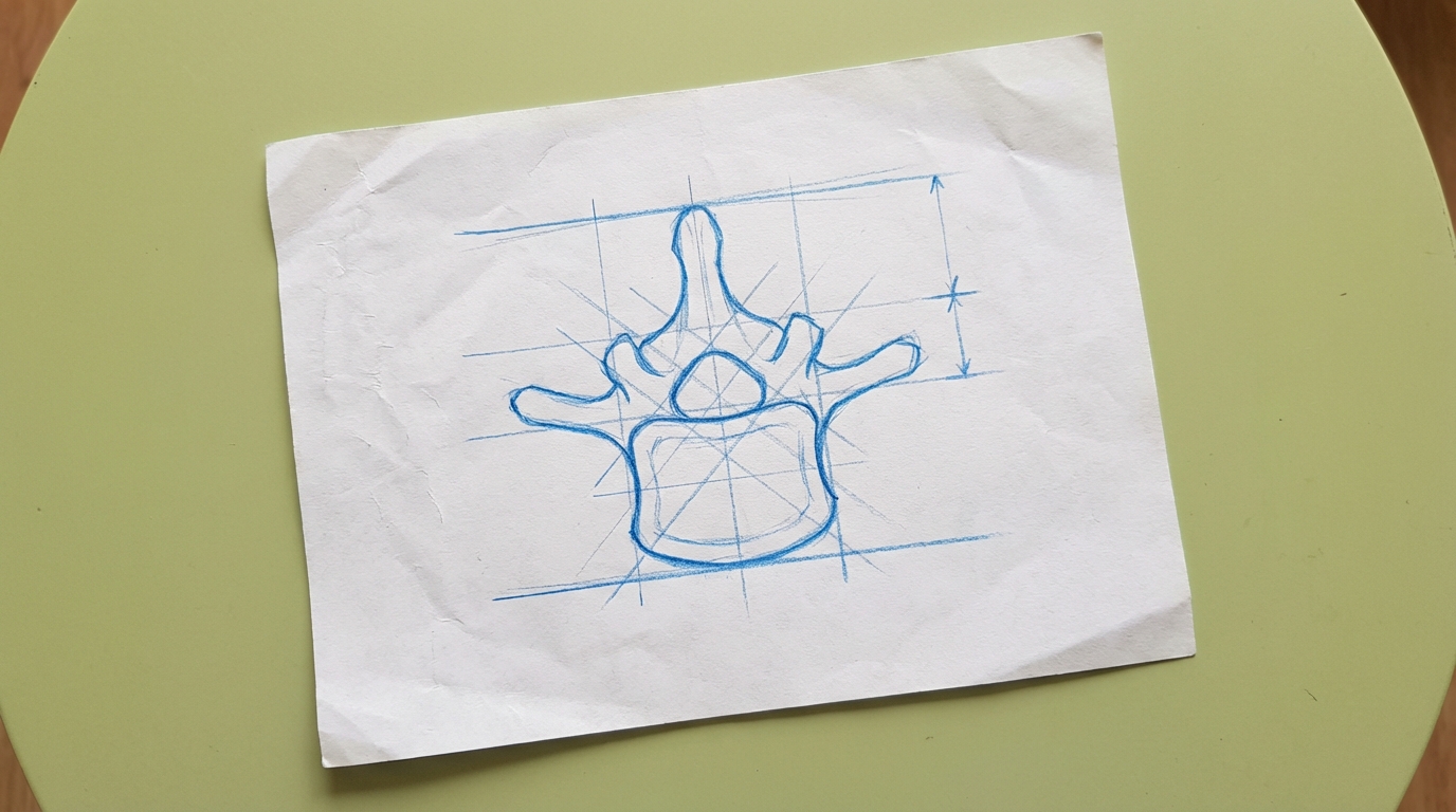
To tailor the procedure to an individual's anatomy, doctors need a way to identify specific landmarks on medical images. This study aimed to find such landmarks, called "fluoroscopic biomarkers," that are visible on a type of real-time X-ray called fluoroscopy. The goal was to establish a clear connection between the three-dimensional (3D) shape of a person's vertebrae and how those bones appear on a two-dimensional fluoroscopy image. This pilot study, which measures and analyzes the form of the bones (a morphometric study), used 3D modeling to find visual cues that could help guide the placement of the treatment instrument.
The researchers worked with 60 sets of human lower back bones, which included the lumbar vertebrae and the sacrum (the large bone at the base of the spine). They photographed these bones and used the images to create detailed 3D digital models. They also captured fluoroscopy images of the actual bones. Using computer software, they then simulated fluoroscopy images from the 3D models. This allowed them to directly compare the 3D bone structure with its corresponding 2D X-ray appearance.
Within these 3D models, the researchers virtually placed a thin tube called a cannula in the ideal position for the LMBRFN procedure. This position was parallel to a specific part of each vertebra known as the superior articular process. They then analyzed the angle of this virtual cannula. Placements were classified as "parasagittal" if the angle was less than 15 degrees, and "traditional" if the angle was greater than 15 degrees.
Out of 118 simulated cannula placements, 85 (or 72.0%) were classified as parasagittal, while 33 (or 28.0%) were classified as traditional. By visually comparing the simulated X-rays of the two groups, the researchers identified a potential fluoroscopic biomarker that appeared to differ between them. This visual marker was present in 70 of the 85 cases (82.0%) that required the parasagittal approach. In contrast, for the 33 cases that required the traditional approach, the same biomarker was present in only 10 cases (30.0%).
This morphometric analysis demonstrates that it is possible to use 3D modeling and simulated fluoroscopy to identify potential visual biomarkers for medical procedures. Further clinical studies with patients and additional anatomical measurement studies are needed to create and confirm robust fluoroscopic biomarkers. Developing these tools could help move the field of interventional pain medicine toward a more personalized approach, where treatments are based on a patient's specific anatomy.